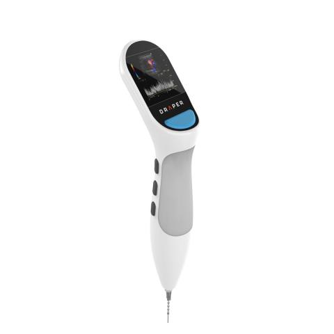
Spectroscopy-Equipped Biopsy Device Could Make Cancer Diagnosis Faster and More Effective
CAMBRIDGE, MA—Thousands of biopsies are performed every year and yet interventional radiologists are typically unaware, until days later, of how many cells have been collected, whether those cells are the suspicious cells that are the target of the biopsy and whether enough cells were collected to detect malignancies.
Draper’s bioengineers are developing an alternative approach. By equipping a fine needle aspiration biopsy syringe with micro-electrical impedance spectroscopy (μEIS) cellular analysis, they are creating a way to enable clinicians to improve their ability to count and classify cancer cells during surgical procedures—two of the major challenges for FNA analysis.
Fine-needle aspiration biopsy is a diagnostic procedure where a needle is inserted into the body, and a small amount of tissue is removed for examination under a microscope. Draper’s micro-EIS system uses the varying electrical properties of human tissue to categorize cellular structures and thereby detect cells whose properties are suspicious and may be cancerous.
Andrew Berlin, distinguished member of the technical staff at Draper, said the new system is an important step in building an FNA biopsy system that can analyze small volumes of patient-derived samples in minutes, not hours or days. “Diagnostic workflows in oncology are underpinned by the need to acquire sufficient cells via biopsy, and to determine which cells in the acquired biopsy are malignant. Draper’s micro-EIS system is uniquely designed to improve a clinician’s ability to count and classify cancer cells while the biopsy procedure is underway at the point of care.”
The new system is described in a paper by Berlin and others presented at the IEEE Conference on Micro and Nanotechnology in Medicine. The authors believe patients could benefit as much as clinicians from the new device, saying, “Rapid sample characterization at the point of care has the potential to reduce the number of biopsy samples that must be acquired, thereby reducing anesthesia duration and potential complications such as bleeding.” The authors added that the system could potentially reduce the number of return visits to repeat biopsy procedures.
Anthony Coston, co-author of the paper and head of medical devices at Draper, said the micro-EIS system is part a movement in biopsy sample analysis that is increasingly shifting towards molecular, rather than multi-cellular, morphological methods. “The system will combine techniques for signature recognition of suspicious cells with downstream molecular analysis to enable the micro-EIS system to characterize a wide range of patient-derived fine needle aspirates for precise, personalized cancer diagnosis right at the point of care. The capabilities required to fully develop this novel medical technology are uniquely Draper,” Coston said.
In tests, a benchtop version of the micro-EIS system distinguished dissociated tumor cells in a sample consisting of red blood cell (RBCs) and peripheral blood mononucleated cells (PBMCs). The micro-EIS system was able to distinguish dissociated tumor cells from normal cells for five cancer types: lung, thyroid, breast, ovarian and kidney cancer. Draper’s device also showed it could make these distinctions in a label-free manner, thereby opening the possibility of integration into standard clinical workflows at the point of care.
Authors are Berlin, Coston and Salil P. Desai, founder and chief executive officer of Phenomyx.
Draper’s micro-EIS system was developed by Draper’s Biomedical Solutions area, which is composed of three areas: Human Organ Systems, Precision Medicine and Biomedical Devices.
Released February 26, 2019

